

 Send Inquiry
ConXport Fluorescent Research Microscope
ConXport Fluorescent Research Microscope Specification
- Features
- Ergonomic design, precise optical alignment, anti-fungal coating on optics, sturdy metallic frame for durability
- Focus System
- Coaxial coarse and fine focus mechanism
- View Head
- Trinocular, Seidentopf type
- Spare Parts
- Spare bulbs, fuse, dust cover, immersion oil
- Theory
- Fluorescent microscopes utilize high-intensity light sources to excite fluorescent stains in specimens, emitting light for detailed observation of biological structures.
- Drawtube
- Trinocular
- Sensor
- Integrated digital camera sensor (optional attachment)
- Resolution
- Up to 1920 x 1080 pixels (camera dependent)
- Interface
- USB 2.0/3.0, HDMI output for camera, PC compatible
- Frame Rate
- 30 fps (with camera module)
- Focal Distance
- Adjustable based on objective used
- Magnification
- 40x - 1600x (objective and eyepiece combination)
- Dimensions
- Approx. 430 mm x 250 mm x 400 mm
- Focus Range
- Coarse and Fine focus with graduated knobs
- Eyepieces
- Paired 10x Wide Field (WF) eyepieces
- Eyepiece Tube
- Inclined at 30, 360 rotatable
- Illumination
- High intensity LED light with fluorescence attachment (Blue, Green filters)
- Coarse Adjustment Range
- 30mm
- Fine Adjustment Range
- 0.002mm
- Working Stage
- Mechanical stage 140 x 140 mm, XY movement 75 x 50 mm
- Still Image Capture Resolution
- HD 1920x1080 pixels
- Video Capture Resolution
- Full HD (camera dependent)
- Image Format
- JPG, BMP (when digital camera is attached)
- Interpupillary Distance
- 55-75 mm adjustable
- Objective Achromatic
- 4x, 10x, 40x (spring), 100x oil immersion (spring)
- Condenser
- Abbe condenser N.A. 1.25 with iris diaphragm and filter holder
- Light Source
- LED with selectable fluorescence excitation
- Microscope Body Material
- Alloy metal, anti-vibration integrated base
- Camera Mounting
- C-mount adapter provided for digital camera
- Packaging
- Supplied in thermocol molded box
- Protection
- Supplied with dust cover and anti-static treatment
- Stage Movement
- X-Y coaxial mechanical movement, smooth and precise
- Application Areas
- Suitable for biology, cytology, microbiology, pathology, fluorescence research and teaching laboratories
- Nosepiece
- Backward quadruple nosepiece
- Power Supply
- AC 220V/110V, 50/60Hz switchable
- Stage Features
- Double layer mechanical stage with low-position controls for easy operation
- Weight
- Approx. 10 kg
- Filter Set
- Standard Blue (Excitation 395-415nm, Emission 460nm) & Green (Excitation 510-550nm, Emission 590nm) filters included
ConXport Fluorescent Research Microscope Trade Information
- FOB Port
- Delhi
- Payment Terms
- Letter of Credit (L/C), Telegraphic Transfer (T/T), Paypal
- Supply Ability
- 15 Per Week
- Delivery Time
- 6-7 Days
- Sample Available
- Yes
- Sample Policy
- If order is confirmed we will reimburse the sample cost
- Main Export Market(s)
- Australia, South America, Eastern Europe, Western Europe, Middle East, Africa, Central America, Asia, North America
- Main Domestic Market
- All India
- Certifications
- ISO,CE,FDA,WHO-GMP
About ConXport Fluorescent Research Microscope
SPECIFICATIONS:
|
Focus: |
Upright microscope with course and fine focus knobs. Built-in Eco switch & possible to upgrade. |
|
Eyepieces: |
10X magnification, with diopter adjustment facility with field of view of 22mm or higher. |
|
Nosepiece: |
Septuple nosepiece with slots for DIC/Polarizing Attachments |
|
Objectives: |
The following objectives suitable for bright field, phase contrast & fluorescence microscopy should be quoted: 1. Plan Achromat 10X 2. Plan Achromat 20X 3. Universal Plan fluorite 40X 4. Universal Plan fluorite 60X 5. Universal Plan fluorite 100 X Oil |
|
Applications: |
The system is equipped with bright field & fluorescence imaging application techniques |
|
Observation Tube: |
Trinocular with observation optical path: 0(Binocular)/100 (Video Port), 100(Binocular)/ 0(Video Port) & 20(Binocular)/ 80(Video Port) Field Number 22mm |
|
Optical System: |
Universal Infinity corrected optical system |
|
Stage: |
Mechanical stage with ceramic coating, with double slide holding capacity |
|
Condenser: |
Condenser for Bright field applications NA 1.1 |
|
Epi-Fluorescence Attachment: |
Epi-fluorescence with Mercury/Metal Halide illuminator of 130W with life-time of 2000 hrs or more. Fluorescence turret with option for at least 5 filter Cubes. It give uniform fluorescence illumination |
|
Fluorescence Filters: |
All the filters have interference type with hard coating for better transmittance/ reflectance. 1. Narrow Band Pass filter for DAPI/Hoechst 2. Narrow Band Pass filter for FITC/GFP 3. Narrow Band Pass filter for TRITC/Rhoda mine/Alexa 546 |
|
Electronics: |
Universal input 110V-240V AC, 50/60 Hz |
|
Illumination: |
Built-in Koehler illumination for transmitted light. Built-in 3 filters (Blue filter, 2 Neutral Density filters). At least 12 V 100W Halogen bulbs (pre-centered) |
|
Optional Accessories: |
Digital USB Cameras (1.3MP,3MP, 5MP, 14MP) with in-built adaptors, Digital Cameras and DSLR Cameras with adaptors, UPS |
Advanced Optics for Detailed Imaging
The microscope offers paired wide field 10x eyepieces and achromatic objectives (4x, 10x, 40x spring, 100x oil immersion spring). Coupled with a Seidentopf trinocular view head and integrated camera compatibility, users enjoy bright, sharp, high-contrast fluorescence images with 40x1600x total magnification.
Precise, User-Friendly Operation
A double layer mechanical stage with low-position controls allows for smooth and accurate sample navigation, while coaxial coarse and fine focusing knobs (30mm/0.002mm ranges) facilitate rapid yet delicate specimen adjustment. The anti-vibration base and ergonomic design ensure comfortable, stable, and fatigue-free operation during lengthy observation sessions.
Flexible Digital Documentation
With a C-mount adapter for digital cameras (optional camera up to 1920x1080 HD), users can capture and store high-quality still images and videos. The systems USB 2.0/3.0 and HDMI outputs provide compatibility with PCs and displays, supporting convenient sharing and analysis of research findings.
FAQs of ConXport Fluorescent Research Microscope:
Q: How do I operate the mechanical stage on the ConXport Fluorescent Research Microscope?
A: The microscope features an X-Y coaxial mechanical stage with low-position controls for precise and comfortable navigation. Simply use the smooth-moving knobs to adjust your sample in both axes, allowing for accurate positioning during observation or imaging.Q: What types of filters are included and how do they enhance fluorescence imaging?
A: This microscope includes standard blue and green filter sets (excitation 395415nm/510550nm, emission 460/590nm). These filters match specific fluorophores, enabling selective excitation and emission to produce clear, high-contrast images of stained biological specimens.Q: When should I use the digital camera attachment, and what are its benefits?
A: Attach the optional C-mount digital camera when you need to document, analyze, or share observations. It offers up to 1920x1080 pixel HD resolution, still image and video capture, and easy output via USB or HDMI, making digital archiving and remote collaboration straightforward.Q: Where is this microscope best suited for use?
A: The ConXport Fluorescent Research Microscope is ideal for biology, cytology, microbiology, pathology research, and educational settingsespecially those involving fluorescence techniques. Its versatile design fits seamlessly in research and teaching laboratories.Q: What makes the focusing process accurate on this microscope?
A: The finely graduated coarse (30mm range) and fine (0.002mm range) focus system ensures quick and highly precise adjustments, optimal for focusing on delicate fluorescent details or thick specimens without losing image sharpness.Q: How does the microscope protect against dust and static damage?
A: It comes supplied with a dust cover and is treated for anti-static protection, preserving optical components and ensuring consistent, dependable performance even in challenging laboratory environments.Q: What are the benefits of the ergonomic and anti-fungal design features?
A: The ergonomic layout, including inclined and 360 rotatable eyepiece tubes, reduces user fatigue during extended sessions. Anti-fungal coatings on the optics safeguard against microbial growth, enhancing durability and maintaining clear imaging over time.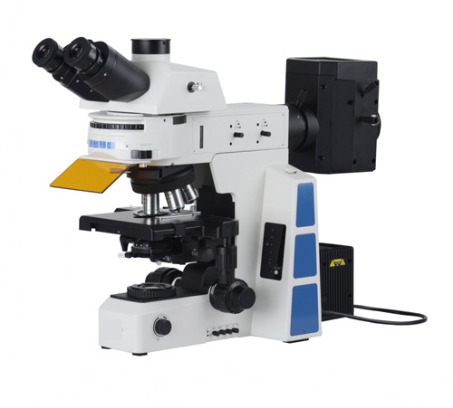
Tell us about your requirement

Price:
Quantity
Select Unit
- 50
- 100
- 200
- 250
- 500
- 1000+
Additional detail
Mobile number
Email

More Products in Microscopes And Projector Category
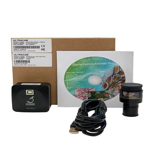
ConXport ULTRACAM Series C-mount USB 2.0 CMOS Camera
Focal Distance : Infinitycorrected
Spare Parts : USB cable, Cmount adapter, installation software
Interface : USB 2.0
Illumination : Supported via microscope illumination
Image Format : JPEG, BMP, PNG, TIFF
Resolution : 5 MP (2592 x 1944 pixels)
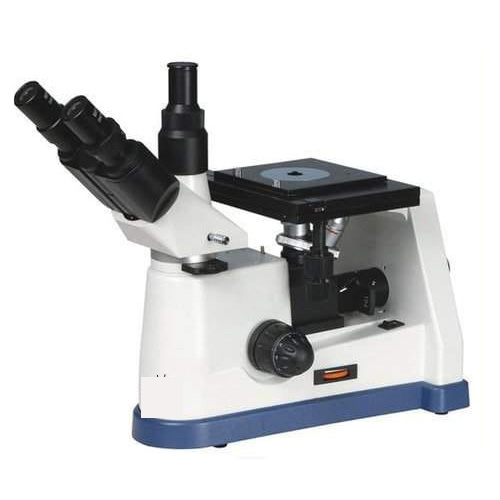
ConXport . Inverted Metallurgical Microscope
Focal Distance : 75mm (standard working distance depending on objective)
Spare Parts : Halogen lamp, fuse, dust cover, stage clips, eye guard, instruction manual
Interface : Trinocular tube for camera attachment
Illumination : 6V 20W Halogen Lamp, Variable intensity
Image Format : Dependent on camera attached (trinocular port), typically supports JPEG/PNG if camera used
Resolution : Optical resolution depends on objective and eyepiece combination
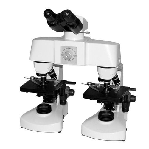
ConXport Malaria Detection Microscope
Focal Distance : Approx. 545 mm (depending on objective lens used).
Spare Parts : Supplied with spare bulb, fuse, and optional immersion oil bottle.
Interface : Direct visual (no digital output); optional camera port for digital models.
Illumination : Builtin LED illumination with adjustable intensity; mirror for natural light.
Image Format : NA (Standard Optical Model); optional camera offers JPEG format stills.
Resolution : Optical, dependent on eyepiece and objective combination.
ConXport HDMI ULTRAHDCMOS720200 C-mount HDMI CMOS Camera
Focal Distance : Adjustable
Spare Parts : Power adaptor, HDMI cable
Interface : HDMI
Illumination : Dependent on microscope illumination
Image Format : JPEG, BMP
Resolution : 1920 x 1080 (Full HD)
|
CONTEMPORARY EXPORT INDUSTRY
GST : 06AJMPT4011D1ZG
GST : 06AJMPT4011D1ZG
- B No. 4353, Main Road, Near Geeta Gopal Temple,Ambala Cantt - 133001, Haryana, India
- Phone :08045815886
- Mr Akhil Trehan (Proprietor)
- Mobile :08045815886
- Send Inquiry

CONTEMPORARY EXPORT INDUSTRY
All Rights Reserved.(Terms of Use)
Developed and Managed by Infocom Network Private Limited.
Developed and Managed by Infocom Network Private Limited.

