

 Send Inquiry
ConXport Malaria Detection Microscope
ConXport Malaria Detection Microscope Specification
- View Head
- Monocular, 360 rotatable, inclined for comfort.
- Features
- Robust metal construction, anti-fungal optics, portable handle, vibration-free base.
- Spare Parts
- Supplied with spare bulb, fuse, and optional immersion oil bottle.
- Focus System
- Coaxial coarse and fine focusing system with sturdy knobs.
- Theory
- Used for rapid and accurate malaria parasite detection in blood smears through microscopy.
- Drawtube
- Monocular, 360 rotatable.
- Sensor
- NA (Optical Microscope, no digital sensor included by default).
- Resolution
- Optical, dependent on eyepiece and objective combination.
- Interface
- Direct visual (no digital output); optional camera port for digital models.
- Frame Rate
- NA (No built-in camera in standard unit).
- Focal Distance
- Approx. 5-45 mm (depending on objective lens used).
- Magnification
- 40X to 1000X (with Achromatic objectives and 10X eyepiece).
- Dimensions
- Approx. 340 x 200 x 130 mm.
- Focus Range
- Coarse: 15mm; Fine: 0.002mm precision.
- Eyepieces
- Pair of WF10X and WF15X included.
- Eyepiece Tube
- Straight monocular tube, inclined at 45 for viewing comfort.
- Illumination
- Built-in LED illumination with adjustable intensity; mirror for natural light.
- Coarse Adjustment Range
- Up to 15mm.
- Fine Adjustment Range
- Up to 0.002mm per division.
- Working Stage
- Mechanical stage 125 x 115 mm with graduated scale and two slide clips.
- Still Image Capture Resolution
- NA (Available with optional camera accessory).
- Video Capture Resolution
- NA (Available with optional camera accessory).
- Image Format
- NA (Standard Optical Model); optional camera offers JPEG format stills.
- Interpupillary Distance
- NA (Single eyepiece model).
- Objective Achromatic
- Achromatic objectives 4X, 10X, 40X(S), 100X(S, Oil).
- Condenser
- NA 1.25 Abbe condenser with iris diaphragm and filter holder.
- Light Source
- LED light with variable intensity or plano-concave mirror.
- Body Material
- Sturdy die-cast aluminum frame with anti-rust coating.
- Dust Cover
- Supplied with protective dust cover for storage.
- Stage Clips
- Twin stage clips provided for slide placement.
- Stage Movement
- X-Y mechanical stage with smooth movement and graduated readings.
- Power Supply
- AC 220V/50Hz or AA battery operated for portability.
- Optics
- Anti-fungal treated lens system for tropical environments.
- Mirror Type
- Plano-concave mirror fitted for use under natural light conditions.
- Safety Features
- Shock-proof base and anti-slip feet.
ConXport Malaria Detection Microscope Trade Information
- FOB Port
- Delhi
- Supply Ability
- 15 Per Week
- Delivery Time
- 6-7 Days
- Sample Available
- Yes
- Sample Policy
- If order is confirmed we will reimburse the sample cost
- Main Export Market(s)
- Western Europe, Australia, North America, South America, Eastern Europe, Middle East, Central America, Asia, Africa
- Main Domestic Market
- All India
- Certifications
- ISO,CE,FDA,WHO-GMP
About ConXport Malaria Detection Microscope
SPECIFICATIONS:
Upgradeyour light microscope into an LED/ Halogen fluorescent microscope at a fractionof the cost - use in the same way as a traditional fluorescent microscope. The LensAdvance fluorescent microscope attachment is a cutting-edge attachment designedto provide the many benefits of LED fluorescence microscopy to any lightmicroscope. LED fluorescence microscopy offers improved detection of manyinfectious diseases. Digital video and image capture is also available.
Features of Lens Advance System:
Convertsany conventional laboratory microscope into a low cost fluorescent microscope
Lowcost alternative to purchasing a traditional fluorescent microscope
Canbe used in the same way as any traditional fluorescent microscope
Simplyattach the Lens Advance to the objectiveposition of your existing light microscope
Ideal for performing any fluorescent microscopy techniques in the field
Advance can also perform:
Accuratelyidentify species - including filarial in blood (using the Q BC Malaria Tube)
Detect Malaria, TB, Trypanosomes and other blood-borne parasites
Quantify the level of parasitaemia
Perform tests for:
Chlamydia,ANA, PCP, CMV: Acid-fast bacilli in sputum, urine
Quickcell count with two part differential count
Plateletcounts for monitoring dengue patients
Identifiesthe need for, and the most appropriate treatment
Canbe used in the laboratory or the field
| Objective | DIN Fluorescent Lens Long working Distance Achromatic objective 62X (N.A. 0.85, W.D. 4.1mm), antifungal and anti reflection coated. |
| Illumination | External illumination 3W Cool Light LED 3.3V with variable control. Up to 100,000 hours of LED life. |
| Electronics | Input 220V AC, 50/60 Hz. |
| Battery back-up | OPTIONAL Chargeable battery back-up 6V, 1.2Ah, Approx. 8 hrs, if fully charge. |
|
| |
| Optional Accessories | 30 Trinocular Head, Digital USB Cameras (1.3MP,3MP, 5MP, 14MP) with in-built adaptors and DSLR Cameras with adaptors, MCFL-550, High Performance 5.0 MP for Fluorescence. |
Optimal Malaria Detection
Rapid and accurate identification of malaria parasites is possible thanks to high-quality achromatic objectives, anti-fungal lenses, and precise focusing mechanisms. The microscopes graduated mechanic stage ensures exact slide movement, essential for thorough examination of blood smears, enabling reliable and repeatable findings vital for effective malaria management.
Portable and Versatile Design
Whether in a clinic or remote location, this microscope adapts to varied settings. An AC 220V power supply or AA batteries deliver flexibility, while the compact, sturdy frame and provided dust cover support safe transport and storage. The instruments shock-proof base and anti-slip feet guarantee stable operation wherever malaria detection is required.
Robust Build for Demanding Settings
Engineered with a die-cast aluminum frame and sealed anti-fungal optics, the microscope is ideal for tropical regions susceptible to harsh conditions. Twin stage clips secure slides during examination while adjustable LED illumination or natural light via plano-concave mirror ensures visual clarityproviding confidence in performance, durability, and hygiene.
FAQs of ConXport Malaria Detection Microscope:
Q: How does the ConXport Malaria Detection Microscope facilitate accurate malaria diagnosis?
A: This microscope uses achromatic objectives, precise coaxial coarse and fine focusing, and a smoothly graduated mechanical stage to allow detailed and systematic examination of blood smears, aiding in the identification of malaria parasites with high reliability.Q: What safety features are included for field and clinic use?
A: It features a shock-proof base, anti-slip feet, robust die-cast frame, and anti-rust coating, offering protection against physical and environmental hazards. The supplied dust cover ensures protection against contamination during storage.Q: When and where can this microscope be used effectively?
A: Its suitable for use in clinics, laboratories, research facilities, and remote field locations, owing to its battery operation option, portable design, and durable construction which withstands frequent transport and tropical conditions.Q: What is the process for examining blood smears with this microscope?
A: Prepare and mount blood smear slides using the twin stage clips, select desired objectives for magnification (40X to 1000X), adjust LED illumination or use natural light with the mirror, and employ the mechanical stage for systematic scan. Fine and coarse focus adjustments enhance image clarity for parasite detection.Q: How does the anti-fungal lens system benefit users in tropical environments?
A: The anti-fungal treated optics prevent lens degradation and fungal growth, ensuring prolonged clarity and reliable results particularly in humid, tropical climates where standard optics may be compromised.Q: Is it possible to capture images from this microscope?
A: The standard model provides direct visual observation through the monocular head. For digital documentation, an optional camera accessory can be added, enabling still images in JPEG format.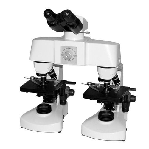
Tell us about your requirement

Price:
Quantity
Select Unit
- 50
- 100
- 200
- 250
- 500
- 1000+
Additional detail
Mobile number
Email

More Products in Microscopes And Projector Category

ConXport Digital Microscope ARGLabs50
Drawtube : Trinocular
Working Stage : Doublelayer mechanical stage, size 140 mm x 140 mm, travel 76 mm x 50 mm
Focal Distance : 8 mm
Magnification : 40x 1000x (4x, 10x, 40x, 100x objectives)
Light Source : LED, variable intensity
Eyepieces : Wide field WF10x/20mm
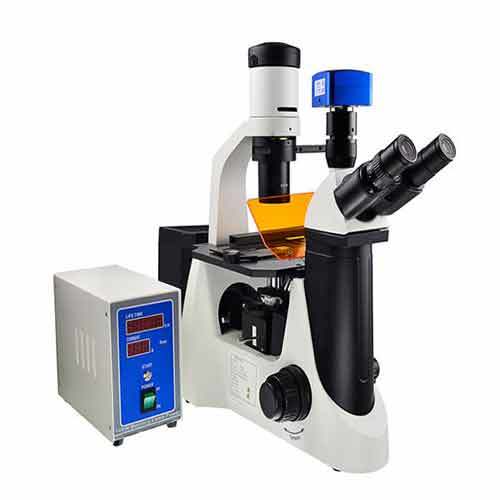
ConXport . Fluorescent Research Microscope
Drawtube : Other, Trinocular, 30 inclined, rotatable 360.
Working Stage : Double layer mechanical stage, size approx. 140 x 140 mm, moveable range 75 x 55 mm.
Focal Distance : 45 mm (standard parfocal distance).
Magnification : 40x 1000x (with standard objectives and eyepieces).
Light Source : High intensity LED or mercury vapor fluorescent illuminator, 5W to 100W depending on configuration.
Eyepieces : Wide field 10x/22mm paired eyepieces.
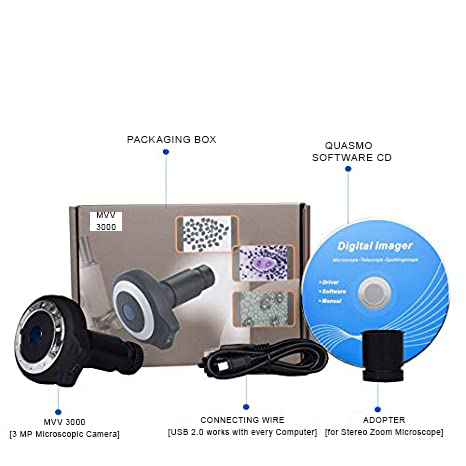
ConXport . CAMERA EYEPIECE
Drawtube : Other, Fits standard 23.2mm microscope eyepiece tubes.
Working Stage : Standard microscope stagecompatible
Focal Distance : Infinitycorrected optics supported
Magnification : 10x eyepiece equivalent
Light Source : Microscopes own illumination system
Eyepieces : Compatible with standard widefield eyepieces
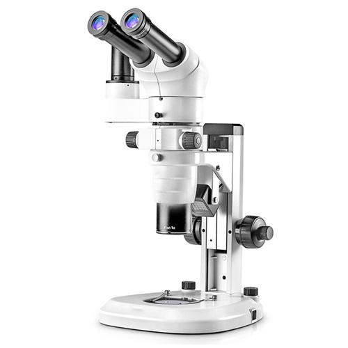
ConXport . Stereo Zoom Microscope
Drawtube : Other, Binocular/Trinocular
Working Stage : Large stage with frosted glass and black & white contrast plates
Focal Distance : 100mm
Magnification : 8x to 50x (zoom)
Light Source : Builtin LED (top and bottom lighting)
Eyepieces : Wide field 10x/20mm paired eyepieces (optional: 15x, 20x)
|
CONTEMPORARY EXPORT INDUSTRY
GST : 06AJMPT4011D1ZG
GST : 06AJMPT4011D1ZG
- B No. 4353, Main Road, Near Geeta Gopal Temple,Ambala Cantt - 133001, Haryana, India
- Phone :08045815886
- Mr Akhil Trehan (Proprietor)
- Mobile :08045815886
- Send Inquiry

CONTEMPORARY EXPORT INDUSTRY
All Rights Reserved.(Terms of Use)
Developed and Managed by Infocom Network Private Limited.
Developed and Managed by Infocom Network Private Limited.

