

 Send Inquiry
ConXport . Slit Lamp (Ultima)
ConXport . Slit Lamp (Ultima) Specification
- Frequency
- 50/60 Hz
- Voltage
- 220 V
- Automation Grade
- Manual
- Capacity
- Slit Lamp offers a magnification range, commonly 10x, 16x, and up to 25x.
- Core Components
- Microscope, illumination system, mechanical base
- Measurement Range
- Microscopic examination up to 16 mm slit length
- Temperature Range
- Ambient room temperature operation
- Feature
- Bright, steady illumination; variable slit width and height; smooth joystick movement
- Model No
- Ultima
- Accuracy
- High precision optical alignment
- Power Source
- AC mains, 220-240V
- Equipment Materials
- High-grade aluminum alloy, coated finish
- Type
- Diagnostic Ophthalmic Equipment
- Usage
- Eye examination, detailed retinal and anterior segment evaluation
- Display Type
- Optical view through binocular microscope
- Dimension (L*W*H)
- Approx. 350 x 420 x 700 mm
- Weight
- Approx. 9-12 kg
- Safety Compliance
- CE marked, ISO certified
- Eyepiece
- 12.5x wide field
- Filter Options
- Cobalt blue, red-free, heat absorbing filters
- Color
- White and Grey Contrasts
- Slit Width Adjustment
- 0 to 14 mm continuously variable
- Objective
- Adjustable aperture, up to 50 mm
- Working Distance
- 80 mm
- Illumination Bulb
- Halogen bulb, 12V/30W
- Head Movement
- Vertical and horizontal adjustment
- Interpupillary Distance
- Adjustable, 55-75 mm
- Application
- Hospital, Clinic, Ophthalmology Practice
- Magnification System
- Three-step rotating drum for variable magnification levels
- Slit Height Adjustment
- 1 to 14 mm
- Base Movement
- Smooth XY movement with precision joystick for positioning
ConXport . Slit Lamp (Ultima) Trade Information
- FOB Port
- Delhi
- Payment Terms
- Letter of Credit (L/C), Telegraphic Transfer (T/T), Paypal
- Supply Ability
- 15 Per Week
- Delivery Time
- 6-7 Days
- Sample Available
- Yes
- Sample Policy
- If order is confirmed we will reimburse the sample cost
- Main Export Market(s)
- Australia, North America, South America, Eastern Europe, Western Europe, Middle East, Africa, Central America, Asia
- Main Domestic Market
- All India
About ConXport . Slit Lamp (Ultima)
Slit Lamps - 4 Models
The slit lamp is essentially a simple and generally under-used piece ofequipment. It consists of an illumination system and a binocular observationsystem, which when correctly aligned will result in a coincidental focus of theslit and microscope.
Illumination system
Basically a short focus projector projecting an image of the illuminated slit apertureon to the eye. This part of the system should be flexible to allow varioussizes and shape of slit beam. Usually a rheostat is incorporated and the lamphouse can be rotated. Neutral density, cobalt blue and red free filters areusually available, and occasionally a diffuser and polarizer
Using the slit lamp
Commence the examination using the 10x eyepieces and the lower poweredobjective i.e. 1x. Use the lowest voltage setting on the transformer. Selectthe longest slit length by means of the appropriate lever (17). Adjust the chinrest by means of control 27 so that the patients eyes are approximately levelwith the black marker on the side of the head rest. Adjust the height of theslit lamp until the slit beam is centred vertically on the patients eye. Focusthe slit beam on the eye by moving the joystick (1) either towards or away fromthe patient. Coarse positioning can be effected without using the microscopebut critical focussing should be carried out whilst viewing through themicroscope. The slit width is varied by rotating either the left hand or righthand knurled control 10. To vary the angle between illumination and microscopeuse one or other of these same controls as handles.
The slit should be set primarily in the vertical position, butany desired inclination can be achieved by means of the ball handle 15 (notchesat 45, 90, 135; stops at 0 and 180). By tripping the latch 11 and tiltingthe slit lamp column, the beam can be introduced from as much as 20 below thehorizontal. This is mainly used for carrying out gonioscopy.
For observation by sclerotic scatter or other dissociated formsof examination the centering screw 13 is loosened, so that the slit image canbe moved away from the centre of the field of observation. The image iscentered again by tightening the screw. Carry out the techniques describedbelow.
|
Specifications - Binocular Microscope |
|||
|
|
Eye Pieces |
: |
10x, 15x |
|
|
Objective |
: |
1x, 1.6x |
|
|
Total Magnification |
: |
10x, 16x and 24x |
|
Illumination Unit |
|||
|
|
Slit Image Rotation |
: |
0 to 180 |
|
|
Tilting Illumination |
: |
Up to 20 |
|
|
Filter Disc |
: |
Cobalt Filter, Green Filter, Yellow Filter Natural Density and Open Aperture |
|
|
Slit Disphragm Disc |
: |
Wedge Shaped Diaphragm Of Infinitely Variable Slit Lengths |
|
|
Halogen Lamp |
: |
12 Volt 50 Watts |
Precision Eye Examination Made Easy
The ConXport Slit Lamp (Ultima) offers ophthalmic professionals precise control for examining ocular structures. Its high-grade optics provide clear, magnified views for intricate assessments of the anterior segment and retina. Intuitive controls and a responsive joystick allow seamless positioning, enabling effortless targeting and adjustment during patient examinations.
Versatile Magnification and Illumination
With a three-step rotating drum, practitioners can select from multiple magnification levels10x, 16x, and 25xtailoring the view based on the clinical requirement. Halogen illumination delivers consistent, bright light, while a range of optical filters (cobalt blue, red-free, heat absorbing) enhances visualization of different ocular features, supporting comprehensive diagnostics.
Ergonomic and Compliant Design
Built with high-grade aluminum and a coated finish, the ConXport Ultima ensures durability and aesthetic appeal. The adjustable interpupillary distance and smooth XY base movement promote operator comfort. Certified CE and ISO standards affirm its safety, making it ideal for hospitals, clinics, and practitioners across India.
FAQs of ConXport . Slit Lamp (Ultima):
Q: How do I adjust the magnification on the ConXport Slit Lamp (Ultima)?
A: The slit lamp features a three-step rotating drum, allowing you to switch smoothly between three magnification levels, typically 10x, 16x, and 25x. Simply rotate the drum to select your desired magnification for optimal visualization during examinations.Q: What eye conditions can be evaluated using this slit lamp?
A: The ConXport Ultima Slit Lamp is designed for comprehensive anterior segment and retinal assessments. It facilitates the diagnosis and evaluation of conditions such as cataracts, corneal abrasions, conjunctivitis, and various retinal disorders.Q: When should the different filter options be used during an examination?
A: Filters enhance ocular structure visibility under specific conditions. Use the cobalt blue filter for fluorescein staining and corneal assessments, the red-free filter to improve vessel contrast, and the heat absorbing filter for prolonged observation without discomfort.Q: What is the process for adjusting the slit width and height?
A: The lamp offers continuously variable slit width (0 to 14 mm) and height (1 to 14 mm), which can be easily adjusted via dedicated controls. This allows the practitioner to customize the light beam according to the area being examined.Q: Where can the ConXport Slit Lamp (Ultima) be utilized?
A: Designed for versatility, it is ideal for use in hospitals, clinics, and ophthalmology practices, supporting a wide range of diagnostic applications in both urban and rural healthcare settings.Q: How does the ergonomic design benefit the user during prolonged use?
A: The adjustable interpupillary distance and smooth, precision XY joystick enable comfortable, fatigue-free examinations. The sturdy yet lightweight aluminum construction facilitates long-term, stable positioning without compromising on operator control.Q: What are the core components included with the ConXport Slit Lamp (Ultima)?
A: The main components include a binocular microscope, a halogen illumination system, a mechanical base with ultra-smooth movement, and essential optical filters, all integrated to deliver accurate and detailed diagnostic results.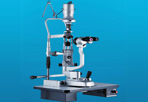
Tell us about your requirement

Price:
Quantity
Select Unit
- 50
- 100
- 200
- 250
- 500
- 1000+
Additional detail
Mobile number
Email

More Products in ENT And Ophthalmic Instruments Category

ConXport Auto Chart Projector
Automation Grade : Automatic
Core Components : Motor, Microprocessor, Optical Lens
Type : Auto Chart Projector
Usage : Eye Examination in Ophthalmology
Equipment Materials : HighQuality Plastic and Metal
Feature : Remote Controlled, Multiple Chart Selections, Programmable
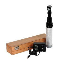
ConXport . Rechargeable Retinoscope Indian
Automation Grade : Manual
Core Components : Retinoscope Head, Rechargeable Handle, Bulb, Mirror
Type : Rechargeable Retinoscope
Usage : Ophthalmic diagnostic examination (Retinoscopy)
Equipment Materials : HighQuality Medical Grade Plastic and Metal
Feature : Streak and Spot Illumination, Rechargeable, Precise Focusing
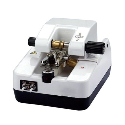
ConXport Optical Lens Groover
Automation Grade : Manual
Core Components : Grooving blade, rotating platform, lens holder
Type : Manual Optical Lens Groover
Usage : Grooving optical lenses for frames
Equipment Materials : Highgrade ABS plastic body
Feature : Precision grooving, manual operation
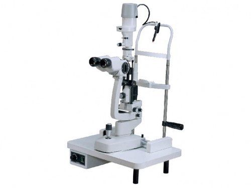
ConXport I. Slit lamp five step
Automation Grade : Manual
Core Components : Microscope, slit lamp, illumination and mechanical base
Type : Fivestep slit lamp
Usage : Ophthalmic examination and diagnosis
Equipment Materials : Highquality metal and optical glass
Feature : Fivestep magnification, precision illumination, ergonomic design
|
CONTEMPORARY EXPORT INDUSTRY
GST : 06AJMPT4011D1ZG
GST : 06AJMPT4011D1ZG
- B No. 4353, Main Road, Near Geeta Gopal Temple,Ambala Cantt - 133001, Haryana, India
- Phone :08045815886
- Mr Akhil Trehan (Proprietor)
- Mobile :08045815886
- Send Inquiry

CONTEMPORARY EXPORT INDUSTRY
All Rights Reserved.(Terms of Use)
Developed and Managed by Infocom Network Private Limited.
Developed and Managed by Infocom Network Private Limited.

