

 Send Inquiry
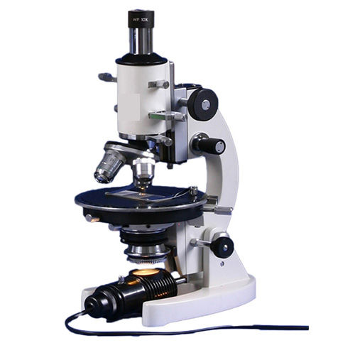
Fluorescent Research Microscope
Fluorescent Research Microscope Specification
- Spare Parts
- Dust cover, power cord, immersion oil, replacement fuse
- Features
- High-contrast fluorescence, ergonomic design, digital imaging, stable stand, anti-fungal optics
- Focus System
- Coaxial coarse & fine focus controls
- View Head
- Trinocular, inclined, 30 & 360 rotatable
- Theory
- Fluorescence microscopy utilizes the property of fluorescence to generate an image and study properties of organic or inorganic substances.
- Drawtube
- Trinocular
- Sensor
- High-resolution CMOS/CCD sensor
- Resolution
- 1920 x 1080 pixels
- Interface
- USB 2.0, HDMI output
- Frame Rate
- 30 fps
- Focal Distance
- 160 mm
- Magnification
- 40X to 1000X (4x, 10x, 40x, 100x objectives)
- Dimensions
- Approx. 400 x 210 x 420 mm
- Focus Range
- Up to 25 mm
- Eyepieces
- Widefield 10x (paired)
- Eyepiece Tube
- Inclined at 30, 360 rotatable
- Illumination
- LED fluorescence illumination, 3W
- Coarse Adjustment Range
- 30 mm
- Fine Adjustment Range
- 0.002 mm per division
- Working Stage
- Mechanical stage 140 mm x 140 mm, moving range 75 mm x 50 mm
- Still Image Capture Resolution
- Up to 5.0 megapixels
- Video Capture Resolution
- Full HD 1080p
- Image Format
- JPG, PNG, BMP
- Interpupillary Distance
- 5575 mm, adjustable
- Objective Achromatic
- 4x, 10x, 40x (spring), 100x oil (spring)
- Condenser
- Abbe condenser NA 1.25 with iris diaphragm and filter holder
- Light Source
- LED (fluorescence), 3W, integrated power supply
- Sample Holder
- Adjustable slide clamps for secure specimen positioning.
- Stage Movement
- Low-position coaxial control for precise XY movement.
- Filter Sets
- Standard blue, green, UV excitation filter blocks included.
- Power Supply
- Universal input: AC 100-240V, 50/60Hz.
- Weight
- Approx. 9 kg net.
- Operating Temperature
- 5C to 40C.
- Fluorescent Attachments
- Removable fluorescence illuminator with multiple filter cubes.
- Camera Port
- Integrated C-mount port for digital camera integration.
- Humidity Control
- Anti-fungal coated optics for humid environments.
- Software
- Image analysis and measurement software included.
About Fluorescent Research Microscope
.Fluorescent Research Microscope
SPECIFICATIONS:
|
Optical system : |
Infinity color corrected optical system |
|
Viewing head: |
30 inclined gemel trinocular head, interpupillary distance: 50mm~76mm; splitting ratio R: T=100:0 or 0:100 |
|
Eyepiece: |
High eye point wide field plan eyepiece PL10X25mm, diopter adjustable. |
|
Objective: |
Plan panchromatic fluorescence objectives (4X/10X/20X/50X/100X) |
|
Nosepiece (with DIC slot): |
Sextuple nosepiece with DIC Slot |
|
Frame: |
Biological frame (transmitted), low-position coaxial coarse and fine adjustment, coarse adjustment distance: 25mm; fine precision: 0.001mm. With coarse adjustment stop and tightness adjustment. Built-in 100-240V_AC50/60Hz wide voltage transformer, intensity adjustable by digital set and reset; built-in transmitted filters LBD/ND6/ND25) |
|
Stage: |
Double layers mechanical stage, size: 187mm X168mm; moving range: 80mm X55mm; precision: 0.1mm; two-way linear drive, tension adjustable |
|
Condenser : |
Swing-out type achromatic condenser (N.A.0.9) |
|
Reflected fluorescence: |
Sextuple reflected fluorescence illuminator with iris field diaphragm and aperture diaphragm, central adjustable; with filter slot and polarizing slot; with fluorescence filters (UV/B/G for option). |
|
Illuminator: |
Digital power controller, wide voltage 100-240VAC |
|
Lamp House: |
12V/100W halogen lamp house for transmitted light, center pre-set, intensity adjustable |
Intuitive Digital Imaging Integration
The microscopes integrated C-mount port makes it simple to connect digital cameras for high-resolution image and video capture. Users can obtain still images up to 5.0 megapixels and Full HD 1080p video, which are saved in common formats such as JPG, PNG, and BMP. Connectivity via USB 2.0 and HDMI output ensures compatibility with various devices, supporting seamless image analysis and documentation in modern laboratory workflows.
Versatile Fluorescence Capabilities
Equipped with standard blue, green, and UV excitation filter blocks alongside removable fluorescence illuminator and multiple filter cubes, this microscope is capable of analyzing a wide array of fluorescent samples. High-contrast fluorescence and stable illumination enable clear observation of specimen details. The ergonomic trinocular view head, inclined and rotatable, allows for comfortable, flexible viewing during extended study sessions.
Precision Focusing and Stage Control
Coaxial coarse and fine focus controls with impressive ranges0.002 mm per division for fine adjustmentsensure exact focusing. The large mechanical stage offers generous space and precise XY movement using low-position coaxial controls. Adjustable slide clamps securely hold samples, while the Abbe condenser with iris diaphragm and filter holder allows optimal light management for clear imaging.
FAQs of Fluorescent Research Microscope:
Q: How do you operate the fluorescence illuminator and switch between filter cubes?
A: The microscope features a removable fluorescence illuminator equipped with multiple filter cubes. To operate, turn on the LED fluorescence light, then select the desired excitation filter (blue, green, or UV) by rotating the filter cube turret. This enables tailored excitation for different fluorescent dyes in your specimens.Q: What is the main benefit of using this microscope for fluorescence studies?
A: Its high-contrast fluorescence capabilities and standard filter sets allow for clear imaging of a variety of fluorescently labeled samples. The microscopes ergonomic design, stable stand, and anti-fungal optics support reliable long-term use, while digital integration ensures efficient data capture and analysis.Q: When should I adjust the interpupillary distance or use the inclined, rotatable trinocular head?
A: Adjust the interpupillary distance (5575 mm) and incline or rotate the trinocular head to ensure comfortable, strain-free viewing during long observation sessions or when sharing the microscope across multiple users. This flexibility optimizes comfort and viewing angles for users of varying preferences.Q: Where can digital images or videos captured by the microscope be accessed or saved?
A: Captured images and videos can be accessed via the integrated camera port, with outputs through USB 2.0 or HDMI. Files in JPG, PNG, or BMP formats can be transferred directly to connected computers or compatible devices for further analysis using the included image measurement software.Q: What environmental conditions are optimal for using this microscope?
A: The device is designed for operation in temperatures ranging from 5C to 40C. Its anti-fungal coated optics make it suitable for humid laboratory environments, ensuring reliable performance and durability in varied climates.Q: How does the microscope ensure specimen stability and precise focusing during observation?
A: The mechanical stage features adjustable slide clamps to securely hold specimens in place, while the coaxial coarse and fine focus controls (with fine adjustment as precise as 0.002 mm per division) enable detailed and stable focusing, minimizing any drift during imaging.Q: What is the process for changing objectives to adjust magnification?
A: The microscope includes a rotating nosepiece fitted with 4x, 10x, 40x (spring), and 100x oil (spring) achromatic objectives. Simply rotate the nosepiece to align the desired objective with the optical path, then refocus as needed to achieve the chosen magnification (from 40X to 1000X).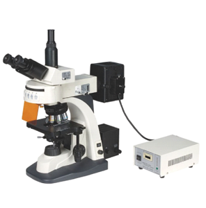
Tell us about your requirement

Price:
Quantity
Select Unit
- 50
- 100
- 200
- 250
- 500
- 1000+
Additional detail
Mobile number
Email

More Products in Microscope Category
Portable Automatic Inclined Binocular
Price 82000 INR / Piece
Minimum Order Quantity : 5 Pieces
Magnification : 40X to 2000X
View Head : Inclined at 45 degrees rotatable 360 degrees
Working Stage : Double layer mechanical stage with XY movement
Theory : Other, Optical Microscopy
PZ-9S Medical Microscope
Magnification : 40X 1000X
View Head : Trinocular, 360 Rotating
Working Stage : Double Layer Mechanical Stage, 140x140 mm
Theory : Other, Medical Microscopy
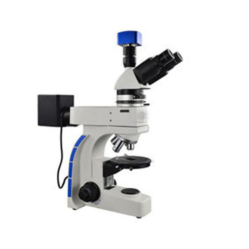
Polarizing Microscope
Magnification : 40x ~ 400x
View Head : Trinocular
Working Stage : Rotatable round mechanical stage
Theory : Other, Polarized light microscopy for birefringent materials analysis
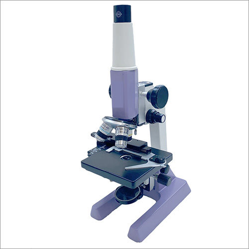
QSP1 Student Polarizing Microscope
Magnification : 40x to 400x (with included objectives and eyepiece)
View Head : Monocular viewing head inclined at 45
Working Stage : Metallic circular stage with 360 rotation
Theory : Other, Polarizing microscope theory for crystalline structures
|
CONTEMPORARY EXPORT INDUSTRY
GST : 06AJMPT4011D1ZG
GST : 06AJMPT4011D1ZG
- B No. 4353, Main Road, Near Geeta Gopal Temple,Ambala Cantt - 133001, Haryana, India
- Phone :08045815886
- Mr Akhil Trehan (Proprietor)
- Mobile :08045815886
- Send Inquiry

CONTEMPORARY EXPORT INDUSTRY
All Rights Reserved.(Terms of Use)
Developed and Managed by Infocom Network Private Limited.
Developed and Managed by Infocom Network Private Limited.

