

 Send Inquiry
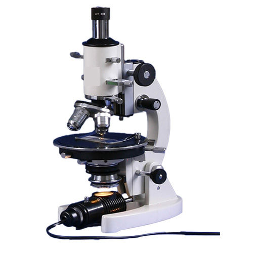
Comparasion Microscope
Price 70000 INR/ Piece
MOQ : 2 Pieces
Comparasion Microscope Specification
- Focus System
- Coaxial coarse and fine focusing
- Spare Parts
- Replacement objectives, eyepiece, immersion oil, dust cover
- View Head
- Dual-view, vertical/horizontal split, 360 rotatable
- Features
- Direct comparison viewing, high-resolution digital imaging, ergonomic design, anti-fungal optic coatings, compatible with measurement software
- Theory
- A Comparison Microscope allows side-by-side observation of two specimens, ideal for detailed comparison such as forensic analysis.
- Drawtube
- Binocular, Siedentopf type
- Sensor
- High-sensitivity CMOS digital imaging sensor
- Resolution
- 1920 x 1080 pixels (Full HD)
- Interface
- USB 2.0 and HDMI outputs
- Frame Rate
- Up to 30 fps
- Focal Distance
- 65 mm (approx.)
- Magnification
- 40x to 1000x (with standard objectives)
- Dimensions
- Approx. 540 mm (L) x 300 mm (W) x 450 mm (H)
- Focus Range
- Coarse and fine adjustments, 30 mm total travel
- Eyepieces
- Wide Field 10x/20mm paired (2 units)
- Eyepiece Tube
- Binocular tube, 45 inclined, 360 rotatable
- Illumination
- Koehler 3W LED illumination with adjustable intensity
- Coarse Adjustment Range
- 24 mm
- Fine Adjustment Range
- 0.002 mm precision
- Working Stage
- Double-layer mechanical stage, 140 mm x 140 mm, X-Y movement 75 mm x 50 mm
- Still Image Capture Resolution
- 5 MP, 8 MP selectable
- Video Capture Resolution
- 1920 x 1080 pixels at 30 fps
- Image Format
- JPG, BMP, PNG, TIFF for still images; AVI/MOV for video
- Interpupillary Distance
- 47 mm - 75 mm adjustable
- Objective Achromatic
- 4x, 10x, 40x (S), 100x (oil) with Plan Achromatic objectives
- Condenser
- Abbe condenser, N.A. 1.25 with iris diaphragm and filter holder
- Light Source
- Integrated, long-life LED module
- Color
- White/Black professional finish
- Stage Type
- Dual synchronized mechanical stages
- Optical Bridge
- Prism-based, high-efficiency light guidance
- Microscope Weight
- 8 Kg (approx)
- Power Supply
- AC 220V / 50Hz
- Packaging
- Sturdy foam-fitted export quality case
- Microscope Body Material
- Die-cast Aluminum alloy
- Supported Operating Systems
- Windows XP/7/8/10, Mac OS (with compatible software)
- Operating Humidity
- 20%-80% RH, non-condensing
- Camera Mount Type
- C-Mount adapter included
About Comparasion Microscope
Comparison Microscope is a specialized type of microscope that allows for simultaneous viewing of two separate samples or objects side-by-side. It consists of two separate microscope bodies connected by an optical bridge, which enables the user to view two samples at once, and make comparisons between them. The microscope typically features high magnification and high-resolution optics, allowing for detailed inspection and analysis of small objects and surfaces. It may also include various illumination options, such as LED or fiber optic lighting, to provide bright and clear images of the samples being examined. Comparison Microscopes are commonly used in forensic science, ballistics, and other fields where side-by-side comparisons are necessary for identification and analysis. They are known for their ability to provide accurate and precise comparisons between two samples, helping to identify similarities or differences that might not be visible with a single microscope.
Precision Optical Comparison for Professionals
Engineered for detailed comparative analysis, this Comparison Microscope features an advanced prism-based optical bridge and dual synchronized stages. Its robust construction and anti-fungal coatings ensure durability and reliability, making it ideal for forensic scientists, researchers, and educators. With an ergonomic design and rotatable dual-view head, the microscope streamlines side-by-side specimen examination for superior accuracy.
Advanced Digital Imaging and Connectivity
Equipped with a high-sensitivity CMOS sensor, the microscope delivers crisp Full HD imaging for both stills and video at up to 30 fps. Connectivity features include USB 2.0 and HDMI outputs, C-Mount camera compatibility, and wide operating system support, ensuring seamless integration into modern digital workflows. Direct image capture and measurements are facilitated through easy-to-use, compatible software.
User-Centric Ergonomics and Comprehensive Features
Operators benefit from adjustable interpupillary distance, coarse and fine focus mechanisms, and a wide field of view for minimal eye strain during extended use. The sturdy mechanical double-layer stage, coaxial adjustments, and built-in Koehler LED illumination with adjustable intensity provide precise control over specimen positioning and lighting, enhancing observation quality and user comfort.
FAQs of Comparasion Microscope:
Q: How does the optical bridge in this comparison microscope enhance specimen analysis?
A: The prism-based optical bridge allows simultaneous, side-by-side observation of two specimens under identical lighting and magnification conditions. This feature is especially valuable for forensic analysis, enabling clear detection of subtle differences or similarities between samples by guiding light efficiently to both optical paths.Q: What operating systems are supported by the microscopes imaging software?
A: The microscope is compatible with Windows operating systems (XP, 7, 8, 10) and Mac OS, provided compatible imaging software is installed. This ensures flexibility for a wide range of users in both educational and professional research facilities.Q: When should I use the 100x oil immersion objective?
A: The 100x oil immersion objective should be used when extremely high magnification and resolution are requiredfor example, in forensic work or laboratory analysis where fine detail needs to be examined. Always apply immersion oil between the slide and the objective lens to maximize image clarity and resolution.Q: Where can this microscope be effectively utilized?
A: This comparison microscope is ideal for forensic laboratories, criminal investigation units, educational institutes, material science labs, and any research environment requiring meticulous comparison of specimens. Its sturdy design and export-quality case also make it suitable for transport between facilities.Q: What is the process for capturing and saving digital images or videos from this microscope?
A: Simply connect the microscope to your computer via USB or HDMI. Use the provided or compatible software to capture still images in JPG, BMP, PNG, or TIFF formats and videos in AVI or MOV formats. You can select image capture resolutions up to 8 MP and video at Full HD (1920 x 1080) and 30 fps.Q: How is optimal illumination achieved during specimen examination?
A: The microscope uses Koehler 3W LED illumination, which provides even light distribution and adjustable intensity. The Abbe condenser with iris diaphragm and filter holder allows fine control over contrast and brightness, ensuring clear and detailed observation across all magnifications.Q: What are the primary benefits of using this comparison microscope in forensic and research applications?
A: Key benefits include precise simultaneous viewing of specimens, high-resolution digital imaging, ergonomic operation reducing user fatigue, adaptable software integration, and long-term durability due to the anti-fungal coated optics and sturdy die-cast aluminum body. These features collectively enhance the reliability and efficiency of forensic, research, and educational workflows.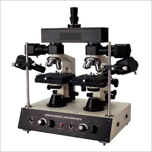
Tell us about your requirement

Price:
Quantity
Select Unit
- 50
- 100
- 200
- 250
- 500
- 1000+
Additional detail
Mobile number
Email

More Products in Microscope Category
Comparison Microscope
Working Stage : Doublelayer mechanical stage, size approx. 145 x 130 mm; movement 70 x 40 mm each side.
Magnification : 40X400X (up to 1000X with optional objectives).
View Head : Dual synchronized optical path for direct specimen comparison.
Theory : Other, Comparison microscopes are designed to compare two specimens side by side via a splitview optical system.
Eyepiece Tube : Interpupillary adjustable 5575mm.
Drawtube : Other, Trinocular drawtube for camera attachment and observation.
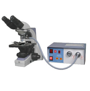
ConXport . Inclined Binocular
Working Stage : Rectangular mechanical stage, size approx. 120 x 140mm, with slide holder.
Magnification : Standard magnification ranges from 40X to 1000X (with objectives: 4X, 10X, 40X, 100X and eyepieces: 10X, 15X).
View Head : Binocular, inclined at 45, rotatable 360.
Theory : Other, Binocular compound microscope designed for educational and laboratory use with an inclined viewing head.
Eyepiece Tube : Binocular tube inclined at 45, adjustable for interpupillary distance.
Drawtube : Other, Binocular, 45 inclined, rotatable 360.
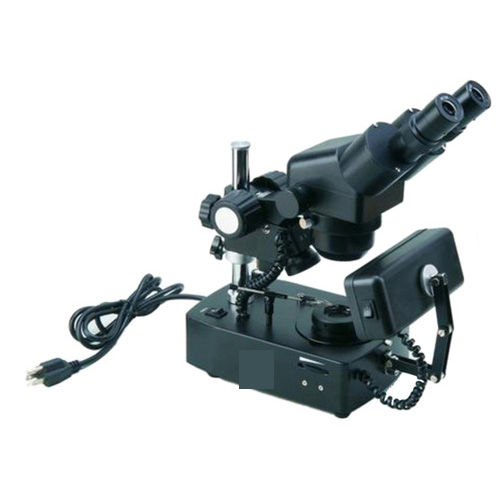
PZ-9 Medical Microscope
Working Stage : Mechanical, 125x115mm
Magnification : 40x 1600x (using various objective and eyepiece combinations)
View Head : Binocular
Theory : Other, Compound optical microscope
Eyepiece Tube : Inclined at 45, rotatable
Drawtube : Binocular
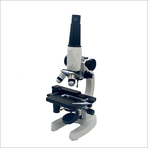
QSP1 Student Polarizing Microscope
Working Stage : Metallic circular stage with 360 rotation
Magnification : 40x to 400x (with included objectives and eyepiece)
View Head : Monocular viewing head inclined at 45
Theory : Other, Polarizing microscope theory for crystalline structures
Eyepiece Tube : One monocular tube inclined at a 45 angle
Drawtube : Other, Standard monocular drawtube
|
CONTEMPORARY EXPORT INDUSTRY
GST : 06AJMPT4011D1ZG
GST : 06AJMPT4011D1ZG
- B No. 4353, Main Road, Near Geeta Gopal Temple,Ambala Cantt - 133001, Haryana, India
- Phone :08045815886
- Mr Akhil Trehan (Proprietor)
- Mobile :08045815886
- Send Inquiry

CONTEMPORARY EXPORT INDUSTRY
All Rights Reserved.(Terms of Use)
Developed and Managed by Infocom Network Private Limited.
Developed and Managed by Infocom Network Private Limited.

