

 Send Inquiry
ConXport Comparison Microscope
ConXport Comparison Microscope Specification
- Focus System
- Coarse and fine coaxial focusing system
- View Head
- Split-field dual viewing with synchronized movement
- Spare Parts
- Spare lamps, eyepieces, objectives, stage clips and dust cover included
- Features
- Precision optical path, modular design for ease of sample comparison, robust metal frame, ergonomic operation for extended use
- Theory
- A comparison microscope is used for side-by-side analysis of specimens, allowing two objects to be viewed simultaneously in a split-field visual format for forensic or analytical examination.
- Drawtube
- Binocular and Trinocular tubes available
- Sensor
- N/A (Eyepiece based visual observation)
- Resolution
- Depends on eyepiece and objective configuration
- Interface
- Eyepiece observation; Trinocular port for camera attachment
- Frame Rate
- Depends on attached camera (if digital option chosen)
- Focal Distance
- 45mm (Standard Working Distance)
- Magnification
- 40x to 400x (Standard); up to 1000x with optional accessories
- Dimensions
- Approx. 480mm x 390mm x 230mm
- Focus Range
- Coarse and Fine adjustment provided
- Eyepieces
- WF10x paired (Interchangeable)
- Eyepiece Tube
- Binocular and Trinocular options, 45 inclined, 360 rotatable
- Illumination
- Koehler illumination with Halogen lamp or built-in LED; adjustable intensity
- Coarse Adjustment Range
- Up to 30mm travel range
- Fine Adjustment Range
- 0.002mm precision movement
- Working Stage
- Mechanical stage, size approx. 140mm x 140mm, dual specimen holding clips
- Still Image Capture Resolution
- Up to 12MP (with suitable camera)
- Video Capture Resolution
- Full HD 1080p supported (with suitable camera)
- Image Format
- Compatible with digital camera for JPEG/RAW output (if camera attached)
- Interpupillary Distance
- 55-75mm adjustable
- Objective Achromatic
- 4x, 10x, 40x, 100x
- Condenser
- Abbe condenser with iris diaphragm, NA 1.25
- Light Source
- Halogen 6V/20W or LED, user selectable
- Accessory Compatibility
- Compatible with most standard digital/CCD/CMOS microscope cameras
- Dust Protection
- Supplied with cover and sealed mechanical parts
- Specimen Holder
- Spring-loaded, dual specimen capacity
- Weight
- Approx. 11.5 kg
- Anti-fungal Coating
- All optical components antifungal treated
- Head Rotation
- Full 360° for convenient sharing and positioning
- Power Supply
- 220V/50Hz AC
- Optical Path Selection
- Left, Right or Split-field via selector switch
- Body Material
- Die-cast aluminum with anti-vibration rubber base
ConXport Comparison Microscope Trade Information
- FOB Port
- Delhi
- Payment Terms
- Letter of Credit (L/C), Telegraphic Transfer (T/T), Paypal
- Supply Ability
- 15 Per Week
- Delivery Time
- 6-7 Days
- Sample Available
- Yes
- Sample Policy
- If order is confirmed we will reimburse the sample cost
- Main Export Market(s)
- Australia, North America, South America, Eastern Europe, Western Europe, Middle East, Central America, Asia, Africa
- Main Domestic Market
- All India
- Certifications
- ISO,CE,FDA,WHO-GMP
About ConXport Comparison Microscope
SPECIFICATIONS:
- HIGH PRECISION COMPARISON MICROSCOPE
- Suitable forensic educational and research,is basically used for forensicscience and police department in large quantity.
- it's also provided these departments such as police training school, bank, tax,print, coinage archaeology and etc.
- Total Magnification 2.4x-200x
- 150W cold light for temperature sensitive objective view
- Light holder with ruler to position light fiber easily
- Video and Camera image output at same time
- Separation Line Adjusting System
- Magnification Correcting System
- Tilt able working stage give different view angle easily
- 5Level Zoom Objective 0.8x,1.2x,3.2x,5x
Innovative Split-Field Viewing
The microscope allows users to view two specimens simultaneously using a split-field format, making it ideal for forensic or analytical comparison work. The synchronized dual view head ensures clear, accurate observation with minimal alignment error, enhancing analytical reliability during side-by-side evaluation.
Precision Built for Stability and Durability
Constructed with die-cast aluminum and supported by an anti-vibration rubber base, the ConXport system guarantees stable performance. The sealed mechanical stage and anti-fungal treated optics ensure the microscope remains dust-free and protected from environmental hazards, even in demanding laboratory conditions.
Flexible Imaging and Ergonomics
This microscope is compatible with standard digital cameras, supporting high-resolution image and video capture for documentation and analysis. The 360 rotatable, 45 inclined head and adjustable interpupillary distance promote user comfort during extended sessions, while the coarse and fine focusing system allows for precise control.
FAQs of ConXport Comparison Microscope:
Q: How do I use the split-field feature of the ConXport Comparison Microscope?
A: To use the split-field viewing, place two specimens in the spring-loaded dual holder. Use the selector switch to toggle between left, right, or split-field modes, enabling simultaneous comparison or focused observation of either specimen.Q: What benefits does the antifungal coating provide?
A: All optical components are antifungal treated, protecting against mold and biological contamination, thereby improving long-term durability and ensuring consistent optical performance even in humid environments.Q: When is it advantageous to rotate the microscope head?
A: The full 360 head rotation allows users to share findings easily, reposition the eyepieces for comfort, and access specimens without moving the entire microscope, which is particularly useful in collaborative or multi-user settings.Q: Where can I attach a digital or CCD/CMOS camera?
A: Digital, CCD, and CMOS cameras can be attached to the trinocular observation port, allowing high-resolution still image and video capture for record-keeping or analysis.Q: What is the process for coarse and fine focusing on this microscope?
A: Adjust your specimen using the coarse focus knob (offering up to 30mm travel) for initial clarity, then use the fine focus control with 0.002mm precision for exacting adjustments, optimizing sharpness and detail for analysis.Q: Which magnification ranges are supported and how can they be expanded?
A: Standard magnification ranges from 40x to 400x, with possible expansion to 1000x using optional accessories like additional objectives and compatible eyepieces.Q: What are the main advantages of the dual specimen holder and mechanical stage?
A: The spring-loaded dual specimen holder and mechanical stage allow secure positioning, easy navigation, and swift comparison, streamlining forensic, biological, or material analyses by keeping samples steady during examination.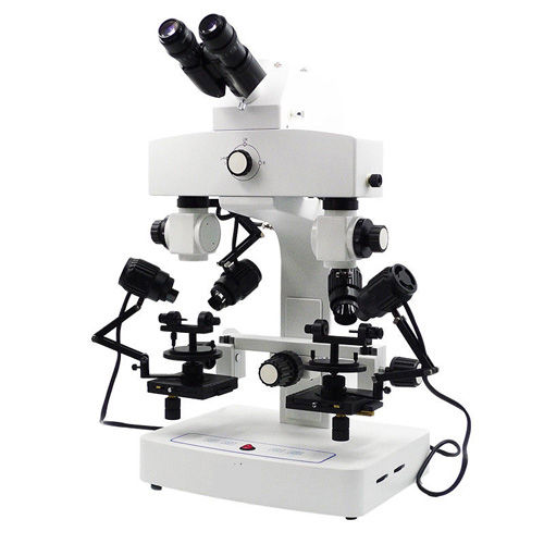
Tell us about your requirement

Price:
Quantity
Select Unit
- 50
- 100
- 200
- 250
- 500
- 1000+
Additional detail
Mobile number
Email

More Products in Microscopes And Projector Category
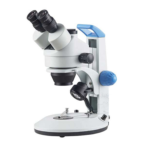
ConXport Stereo Zoom Microscope
Objective Achromatic : Zoom objective 0.7X4.5X, achromatic lenses
Theory : Other, ConXport Stereo Zoom Microscope is designed for high resolution and threedimensional viewing of specimens, suitable for research, clinical, and educational applications.
Focus Range : 50 mm (approx.)
Illumination : Incident and transmitted LED illumination, with adjustable brightness
Eyepieces : WF10X/20 mm (pair)
Image Format : N/A (optical only)
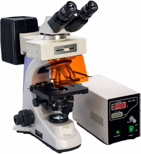
ConXport . Fluorescent Research Microscope
Objective Achromatic : DIN Achromatic 4X, 10X, 40X (spring), 100X (spring, oil)
Theory : Other, A fluorescent research microscope uses fluorescence and phosphorescence techniques for high sensitivity observation, especially useful in biomedical and life science applications.
Focus Range : Coarse: 20 mm; Fine: 0.002 mm precision
Illumination : LED lamp for transmitted light, 100 W highpressure mercury lamp for fluorescence
Eyepieces : Wide field 10X/18mm, paired set
Image Format : JPG, BMP, PNG (with compatible camera/software)
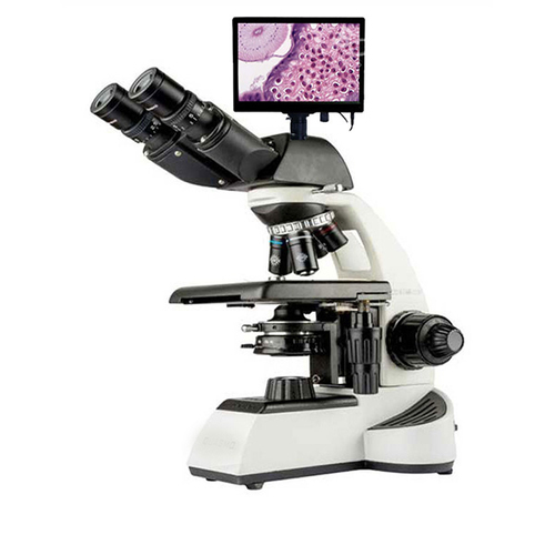
ConXport DIGITAL MICROSCOPE
Objective Achromatic : 4x, 10x, 40x, 100x oil (standard set)
Theory : Other, Digital imaging and display for microscopy applications, allowing direct observation on screen.
Focus Range : Fine and coarse adjustment available
Illumination : Incident and transmitted LED illumination with adjustable brightness
Eyepieces : WF10x (Wide Field Eyepiece)
Image Format : JPEG, BMP, PNG or AVI for video
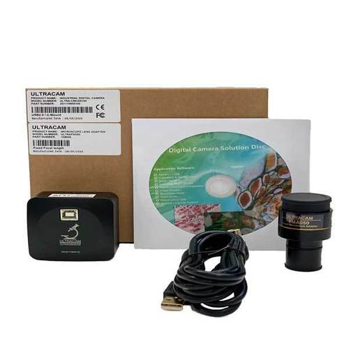
ConXport ULTRACAM Series C-mount USB 2.0 CMOS Camera
Objective Achromatic : Compatible with achromatic objectives (via microscope)
Theory : Other, Digital imaging for microscopy applications using USB connectivity
Focus Range : Fine and coarse adjustment mechanism
Illumination : Supported via microscope illumination
Eyepieces : Compatible via Cmount adapter; eyepiece not included
Image Format : JPEG, BMP, PNG, TIFF
|
CONTEMPORARY EXPORT INDUSTRY
GST : 06AJMPT4011D1ZG
GST : 06AJMPT4011D1ZG
- B No. 4353, Main Road, Near Geeta Gopal Temple,Ambala Cantt - 133001, Haryana, India
- Phone :08045815886
- Mr Akhil Trehan (Proprietor)
- Mobile :08045815886
- Send Inquiry

CONTEMPORARY EXPORT INDUSTRY
All Rights Reserved.(Terms of Use)
Developed and Managed by Infocom Network Private Limited.
Developed and Managed by Infocom Network Private Limited.

