

 Send Inquiry
ConXport . Fluorescent Research Microscope
ConXport . Fluorescent Research Microscope Specification
- View Head
- Trinocular, 45 inclined and 360 rotatable, suitable for photomicrography
- Focus System
- Coaxial coarse & fine focusing mechanism with adjustable tension
- Features
- Equipped with high-efficiency fluorescence attachment, mechanical stage, easy switching between brightfield and fluorescence, robust metal body, anti-fungal optics.
- Spare Parts
- Supplied with spare halogen & mercury lamp, fuses, dust cover, immersion oil, and power cord
- Theory
- A fluorescent research microscope uses fluorescence and phosphorescence techniques for high sensitivity observation, especially useful in biomedical and life science applications.
- Drawtube
- Trinocular; 45 inclined, 360 rotatable
- Sensor
- High sensitivity CMOS/CCD camera compatible (camera not included)
- Resolution
- Up to 1920x1080 (with compatible camera, not included)
- Interface
- USB 2.0 (for camera connectivity)
- Frame Rate
- Up to 30 fps (with compatible camera, not included)
- Focal Distance
- Adjustable, typically 5575 mm interpupillary distance
- Magnification
- 40X 1000X (using 4X, 10X, 40X, 100X objectives and 10X eyepieces)
- Dimensions
- Approx. 455 mm x 285 mm x 395 mm (LxWxH)
- Focus Range
- Coarse: 20 mm; Fine: 0.002 mm precision
- Eyepieces
- Wide field 10X/18mm, paired set
- Eyepiece Tube
- Trinocular head with diopter adjustment on one side
- Illumination
- LED lamp for transmitted light, 100 W high-pressure mercury lamp for fluorescence
- Coarse Adjustment Range
- Up to 20 mm
- Fine Adjustment Range
- 0.002 mm per division
- Working Stage
- Double layer mechanical stage, 140 mm x 135 mm, with vernier scale and low-position coaxial controls; moving range 75 mm x 50 mm
- Still Image Capture Resolution
- Up to 5MP (dependent on camera, not included)
- Video Capture Resolution
- Up to 1920x1080 (dependent on camera, not included)
- Image Format
- JPG, BMP, PNG (with compatible camera/software)
- Interpupillary Distance
- 5575 mm adjustable
- Objective Achromatic
- DIN Achromatic 4X, 10X, 40X (spring), 100X (spring, oil)
- Condenser
- N.A. 1.25 Abbe condenser with iris diaphragm and filter holder
- Light Source
- 100 W mercury lamp (fluorescence); LED transmitted light
- Optical System
- Infinity-Corrected Optical System
- Fluorescence Illuminator
- Built-in, with external lamp house and easy lamp replacement
- Application Areas
- Life sciences, immunofluorescence, cell biology, microbiology, pathology
- Mechanical Stage Travel
- 75 x 50 mm range
- Fluorescence Filter Cubes
- Supplied with B (blue), G (green), and U (UV) cubes for various fluorophores
- Digital Imaging Compatibility
- Supports photo and video documentation with appropriate camera attachment
- Power Supply
- AC 220V/110V ±10%, 50/60Hz
- Eyepiece Field Number
- 18 mm
- Safety Features
- Overheat protection for mercury lamp housing, safety interlock for UV exposure
- Type of Stage Movement
- Low-position, coaxial X-Y controls for smooth movement
- Stage Size
- 140 x 135 mm
ConXport . Fluorescent Research Microscope Trade Information
- FOB Port
- Delhi
- Payment Terms
- Paypal, Letter of Credit (L/C), Telegraphic Transfer (T/T)
- Supply Ability
- 15 Per Week
- Delivery Time
- 6-7 Days
- Sample Available
- Yes
- Sample Policy
- If order is confirmed we will reimburse the sample cost
- Main Export Market(s)
- Australia, North America, South America, Eastern Europe, Western Europe, Middle East, Africa, Central America, Asia
- Main Domestic Market
- All India
- Certifications
- ISO,CE,FDA,WHO-GMP
About ConXport . Fluorescent Research Microscope
FEATURES:
Microscopewith Universal Infinity corrected system for BRIGHTFIELD, FLUORESCENCEAPPLICATIONS AND should be useful to DIC Applications later, Built-in-Kholer 6V/20W Halogen illumination for transmitted light with light preset switch, Lightintensity LED Indicator, built in filters (ND6, ND25 etc) & Hg HBO-100WMercury lamp attachment for Reflected light Fluorescence application and filterset for Blue excitation. EX490nm (blue), EX545nm (Green) exciting light fittersystem, O ordinary light system, Interchangeable Trinocular with inclined 30degree for observation, Ceramic coated scratch proof co-axial stage with leftor right hand low drive X & Y control with torque adjustment, UniversalAbbes condenser for bright field, applications & UV Protection screen.
SPECIFICATIONS:
| Optical System: | Infinite Optical System |
| Viewing Head: | Compensation Free Trinocular Head Inclined at 30° Interpupillary Distance 48-75mm |
| Eyepiece: | Extra Wide Field Eyepiece EW10 x 20 |
| Nosepiece: | Quadruple Nosepiece |
| Objective: | Objective finite Plan Achromatic Objective 4 , 10, 40, 100 |
| Condenser: | Abbe's Condenser NA 0.9/ 1.25 |
| Focusing: | Coaxial Coarse & Fine Adjustment, fine-focusing scale value 0.002mm |
| Stage: | Double Layers Mechanical Stage 180 x 160/ 80x 50 mm |
| Kohler Illumination: | Transmission illumination |
| Lamp: | Halogen Bulb 12V/100W-AC85V-230V |
| Fluorescent | B Wave band |
| Optional | Eyepiece: WF15X ,WF20X |
High-Sensitivity Fluorescence Microscopy
This microscope uses fluorescence and phosphorescence techniques for enhanced detection of labeled specimens. The fluorescence attachment and built-in illuminator allow observation of a variety of fluorophores under B, G, and U filter cubes. Such sensitivity makes it highly effective for biomedical applications, immunofluorescence, and cell studies.
Ergonomic Design and Smooth Operation
The trinocular head is inclined at 45 and rotates 360, supporting both comfortable viewing and photomicrography. The low-position coaxial stage controls ensure smooth and precise movement, while coarse and fine focusing systems provide excellent control for high-resolution imaging. The mechanical stage accommodates a wide range of specimen sizes.
Digital Documentation Capabilities
Compatible with high-sensitivity CMOS/CCD cameras, the microscope supports digital photo and video documentation with up to 1920x1080 resolution and 30 fps frame rate. Images can be captured in JPG, BMP, or PNG formats for analysis and record-keeping, making it fully equipped for research and presentation requirements.
FAQs of ConXport . Fluorescent Research Microscope:
Q: How does the ConXport Fluorescent Research Microscope enhance fluorescence observation?
A: It utilizes high-efficiency fluorescence filter cubes (B, G, U), built-in mercury lamp illumination, and advanced infinity-corrected optics, enabling sensitive detection and visualization of various fluorophores for detailed cellular and molecular analysis.Q: What is the procedure for switching between brightfield and fluorescence modes?
A: The microscope is equipped with both LED transmitted light for brightfield and a 100 W mercury lamp for fluorescence. Users can easily switch modes using the fluorescence attachment and corresponding filter cubes, enabling seamless transition for diverse imaging needs.Q: When is it beneficial to use the trinocular head and digital imaging features?
A: The trinocular head is ideal when simultaneous observation and photo/video documentation are required. With camera compatibility (CMOS/CCD), users can capture high-resolution images and videos during live sample examination for research, presentation, or record-keeping.Q: Where is this microscope most commonly applied?
A: It is widely used in life sciences laboratories, universities, hospitals, pathology labs, and research centers in applications such as immunofluorescence, cell biology, microbiology, and diagnostic pathology.Q: What safety features are included for user protection?
A: The microscope features overheat protection for the mercury lamp housing and a safety interlock for UV exposure, reducing risk during prolonged fluorescent imaging and lamp replacement.Q: How does the mechanical stage facilitate sample examination?
A: The double layer mechanical stage has a size of 140 x 135 mm and a travel range of 75 x 50 mm, with low-position coaxial X-Y controls and a vernier scale for precise and smooth movement, ensuring accurate specimen positioning during observation.Q: What benefits does digital imaging compatibility offer?
A: Digital imaging allows users to record and analyze images and videos of samples with resolutions up to 1920x1080 and 5MP stills (with appropriate camera). It supports various formats, facilitating research documentation, data sharing, and publication.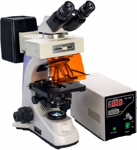
Tell us about your requirement

Price:
Quantity
Select Unit
- 50
- 100
- 200
- 250
- 500
- 1000+
Additional detail
Mobile number
Email

More Products in Microscopes And Projector Category

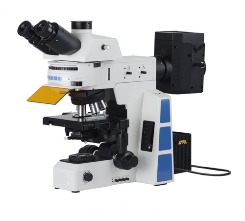
ConXport . Upright Research Fluorescent Microscope
Illumination : LED or Mercury vapor lamp for fluorescence; Koehler Halogen/LED for transmitted light
Focus Range : Coarse: 30 mm; Fine: 0.002 mm precision
Spare Parts : Standard fuse, extra lamp, dust cover, immersion oil
Objective Achromatic : Plan Achromatic Objectives (4X, 10X, 20X, 40X, optional 100X oil)
Sensor : High sensitivity CMOS or CCD (camera optional)
Features : Multichannel fluorescence capability, high stability framework, antimold optics, adjustable brightness and filter turret
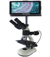
ConXport Digital Microscope iQ
Illumination : Adjustable LED
Focus Range : 030 mm
Spare Parts : Dust cover, immersion oil, power cord, extra fuse
Objective Achromatic : 4x, 10x, 40x, 100x (oil) achromatic objectives
Sensor : CMOS
Features : Snapshot, video recording, measurement software and USB connectivity.
ConXport Fluorescent Research Microscope
Illumination : High intensity LED light with fluorescence attachment (Blue, Green filters)
Focus Range : Coarse and Fine focus with graduated knobs
Spare Parts : Spare bulbs, fuse, dust cover, immersion oil
Objective Achromatic : 4x, 10x, 40x (spring), 100x oil immersion (spring)
Sensor : Integrated digital camera sensor (optional attachment)
Features : Ergonomic design, precise optical alignment, antifungal coating on optics, sturdy metallic frame for durability
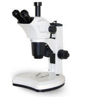
ConXport . Stereo Zoom Microscope
Illumination : Incident and transmitted LED illumination with adjustable brightness.
Focus Range : Coarse and fine focusing mechanism.
Spare Parts : Available: eyepieces, objectives, stage plates, bulbs, fuses.
Objective Achromatic : Zoom objectives 0.7x to 4.5x, achromatic corrected for clear imaging.
Sensor : Not Applicable (Optical observation only)
Features : Ergonomic, continuous zoom, dual LED illumination, antifungal coated optics.
|
CONTEMPORARY EXPORT INDUSTRY
GST : 06AJMPT4011D1ZG
GST : 06AJMPT4011D1ZG
- B No. 4353, Main Road, Near Geeta Gopal Temple,Ambala Cantt - 133001, Haryana, India
- Phone :08045815886
- Mr Akhil Trehan (Proprietor)
- Mobile :08045815886
- Send Inquiry

CONTEMPORARY EXPORT INDUSTRY
All Rights Reserved.(Terms of Use)
Developed and Managed by Infocom Network Private Limited.
Developed and Managed by Infocom Network Private Limited.

