

 Send Inquiry
ConXport . Slit Lamp (Ultima) Stepper Magnification
ConXport . Slit Lamp (Ultima) Stepper Magnification Specification
- Voltage
- 220240 V
- Model No
- Ultima
- Core Components
- Illumination system, binocular microscope, mounting stand
- Power Source
- AC Powered
- Measurement Range
- Slit width: 014 mm, Slit length: 114 mm
- Feature
- Stepper magnification; LED illumination; precision optics
- Frequency
- 50/60 Hz
- Automation Grade
- Manual
- Accuracy
- High precision optical system
- Equipment Materials
- Aluminum alloy, Stainless steel, Optical glass
- Type
- Ophthalmic Examination Equipment
- Usage
- Slit lamp examination for eye diagnosis
- Display Type
- Binocular viewing
- Dimension (L*W*H)
- Approx. 700 x 330 x 760 mm
- Weight
- Approx. 16 kg
- Illumination Spot Size
- 0.1 mm to 14 mm variable
- Eyepiece
- 10x widefield
- Microscope Type
- Galilean converging type
- Focus Travel
- Up to 30 mm
- Interpupillary Distance
- 5278 mm adjustable
- Light Source
- LED lamp, adjustable intensity
- Filter System
- Blue, red-free, heat absorption filters
- Slit Rotation
- 0 to 180 continuous
- Objective Lens
- Superb quality, anti-reflective coating
- Chin Rest Adjustment
- Up to 70 mm
- Base Movement
- XY joystick-controlled; smooth operation
- Magnification Steps
- Three-step (10x, 16x, 25x selectable)
ConXport . Slit Lamp (Ultima) Stepper Magnification Trade Information
- FOB Port
- Delhi
- Payment Terms
- Letter of Credit (L/C), Paypal, Telegraphic Transfer (T/T)
- Supply Ability
- 15 Per Week
- Delivery Time
- 6-7 Days
- Sample Available
- Yes
- Sample Policy
- If order is confirmed we will reimburse the sample cost
- Main Export Market(s)
- Australia, South America, Eastern Europe, Western Europe, Middle East, Africa, Central America, Asia, North America
- Main Domestic Market
- All India
- Certifications
- ISO,CE,FDA,WHO-GMP
About ConXport . Slit Lamp (Ultima) Stepper Magnification
Slit Lamps - 4 Models
The slit lamp is essentially a simple and generally under-used piece ofequipment. It consists of an illumination system and a binocular observationsystem, which when correctly aligned will result in a coincidental focus of theslit and microscope.
Illumination system
Basically a short focus projector projecting an image of the illuminated slitaperture on to the eye. This part of the system should be flexible to allowvarious sizes and shape of slit beam. Usually a rheostat is incorporated andthe lamp house can be rotated. Neutral density, cobalt blue and red freefilters are usually available, and occasionally a diffuser and polarizer
Using the slit lamp
Commence the examination using the 10x eyepieces and the lower poweredobjective i.e. 1x. Use the lowest voltage setting on the transformer. Selectthe longest slit length by means of the appropriate lever (17). Adjust the chinrest by means of control 27 so that the patients eyes are approximately levelwith the black marker on the side of the head rest. Adjust the height of theslit lamp until the slit beam is centred vertically on the patients eye. Focusthe slit beam on the eye by moving the joystick (1) either towards or away fromthe patient. Coarse positioning can be effected without using the microscopebut critical focussing should be carried out whilst viewing through themicroscope. The slit width is varied by rotating either the left hand or righthand knurled control 10. To vary the angle between illumination and microscopeuse one or other of these same controls as handles.
The slit should be set primarily in the vertical position, butany desired inclination can be achieved by means of the ball handle 15 (notchesat 45, 90, 135; stops at 0 and 180). By tripping the latch 11 and tiltingthe slit lamp column, the beam can be introduced from as much as 20 below thehorizontal. This is mainly used for carrying out gonioscopy.
For observation by sclerotic scatter or other dissociated formsof examination the centering screw 13 is loosened, so that the slit image canbe moved away from the centre of the field of observation. The image iscentered again by tightening the screw. Carry out the techniques describedbelow.
S
|
Specifications - Binocular Microscope |
|||
|
|
Viewing Oculars |
: |
Binocular, Galilean Type |
|
|
Magnification Change |
: |
Three Steps Drum Rotation Type Magnification |
|
|
Eye Pieces |
: |
Wide Field 12.5X |
|
|
Objective |
: |
0.6x, 1x, 1.6x |
|
|
Total Magnification |
: |
7.5x, 12.5x, 20x |
|
Illumination Unit |
|||
|
|
Slit Image Rotation |
: |
0 to 180 |
|
|
Tilting Illumination |
: |
5 to 20 |
|
|
Filter Disc |
: |
Cobalt Filter, Red Free, Yellow Filter Natural Density and Open Aperture Diaphragm For Infinitely Variable Slit Lengths. |
|
|
Slit Disphragm Disc |
: |
Six Apertures Of 12, 9, 7, 3 and 0.2mm and a Wedge Shaped Diaphragm Of Infinitely Variable Slit Lengths |
Advanced Magnification for Precise Diagnosis
The Ultima Slit Lamp features a stepper magnification system, giving practitioners three selectable levels (10x, 16x, 25x) for tailored examination. The anti-reflective, high-quality objective lens, combined with widefield eyepieces, ensures clear and detailed visualization, which is critical for accurate diagnosis during ophthalmic procedures.
Ergonomic Design and Smooth Operation
Constructed from durable aluminum alloy and stainless steel, the device offers an ergonomic chin rest adjustment up to 70 mm and a joystick-controlled XY base for fluid positioning. This design streamlines the examination process, maximizes patient comfort, and supports enduring clinical use.
Superior Illumination and Filter System
A high-intensity LED lamp provides adjustable lighting to suit each examination. The integrated filter system offers options like blue, red-free, and heat absorption filters, allowing optimal viewing of various ocular structures. The customizable illumination spot size (0.1 mm to 14 mm) further enhances eye diagnostics.
FAQs of ConXport . Slit Lamp (Ultima) Stepper Magnification:
Q: How does the stepper magnification feature benefit ophthalmic examinations?
A: The three-step magnification (10x, 16x, 25x) allows clinicians to quickly select the optimal zoom level for detailed views of ocular structures, improving diagnostic accuracy and efficiency during eye examinations.Q: What is the process for adjusting the slit lamp to accommodate different patients?
A: Operators can adjust the interpupillary distance (5278 mm), chin rest height (up to 70 mm), and illumination intensity to comfortably fit each patients anatomy and examination needs, using the smooth joystick-controlled base for precise positioning.Q: Where is the ConXport Slit Lamp (Ultima) typically used?
A: This slit lamp is commonly utilized in ophthalmology clinics, hospitals, eye care centers, and vision diagnostic facilities across India and internationally by exporters, manufacturers, suppliers, and traders.Q: When should I use the filter system during examination?
A: Filters like blue, red-free, and heat absorption can be applied as needed: blue for fluorescein staining, red-free for vascular assessment, and heat absorption for protecting ocular tissues during extended exams.Q: What materials are used in the construction of the slit lamp?
A: The Ultima model is manufactured using premium aluminum alloy, stainless steel, and high-grade optical glass, ensuring durability and consistent optical performance.Q: What is the core benefit of the Ultimas LED illumination system?
A: The LED lamp provides adjustable intensity, low heat emission, and long-lasting illumination, making the device energy-efficient and comfortable for both clinicians and patients during extended use.Q: How accurate is the Ultima Slit Lamps optical system?
A: Its precision Galilean converging microscope with anti-reflective coating and high-quality optics delivers sharp, distortion-free images, crucial for high-precision eye diagnosis.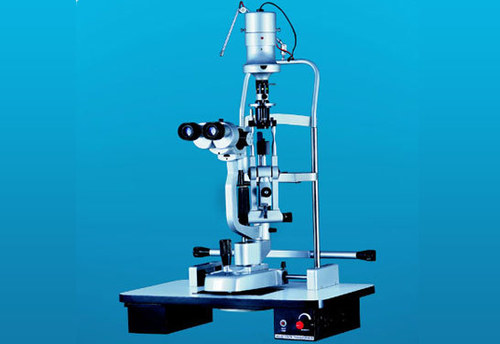
Tell us about your requirement

Price:
Quantity
Select Unit
- 50
- 100
- 200
- 250
- 500
- 1000+
Additional detail
Mobile number
Email

More Products in ENT And Ophthalmic Instruments Category

ConXport Ophthalmic Bipolar Surgical Cautery
Dimension (L*W*H) : Device: 20 cm x 7 cm x 5 cm (approx)
Weight : Approx. 1 kg
Usage : Ophthalmic surgical cauterization
Core Components : Main unit, detachable wire, bipolar forceps, electrodes
Model No : ConXport Ophthalmic Bipolar Cautery
Accuracy : High surgical precision (manufacturer standard)

ConXport Surgical Operating Microscope Cold Light
Dimension (L*W*H) : Base: approx. 650mm x 650mm; Height: adjustable 12001700mm
Weight : Approx. 35 kg (Including Stand)
Usage : Medical Surgery, ENT, Dental, Ophthalmic Procedures
Core Components : Optical Binoculars, LED Cold Light Source, Articulated Stand
Model No : ConXport Surgical Operating Microscope Cold Light
Accuracy : Precision Optical Lenses with MultiLayer Coating
ConXport . I/A, Simcoe Cannulas
Dimension (L*W*H) : Length: 19 mm (typical), Diameter: 22G or 23G
Weight : Approximately 10 g per cannula
Usage : Ophthalmic surgical procedures and anterior chamber irrigation/aspiration
Core Components : Straight or curved cannula with handle, luer lock connector
Model No : ConXport I/A Simcoe Cannula
Accuracy : Precisionformed tip for controlled fluid flow
ConXport Corneal Forceps Toothed Straight
Dimension (L*W*H) : Length: Approx. 110 mm
Weight : Approx. 15g
Usage : Used for grasping and holding corneal tissue during ophthalmic surgery
Core Components : Toothed tips, precision grip handle
Model No : ConXportTCSF110
Accuracy : Fine precision tips for microsurgical procedures
|
CONTEMPORARY EXPORT INDUSTRY
GST : 06AJMPT4011D1ZG
GST : 06AJMPT4011D1ZG
- B No. 4353, Main Road, Near Geeta Gopal Temple,Ambala Cantt - 133001, Haryana, India
- Phone :08045815886
- Mr Akhil Trehan (Proprietor)
- Mobile :08045815886
- Send Inquiry

CONTEMPORARY EXPORT INDUSTRY
All Rights Reserved.(Terms of Use)
Developed and Managed by Infocom Network Private Limited.
Developed and Managed by Infocom Network Private Limited.

